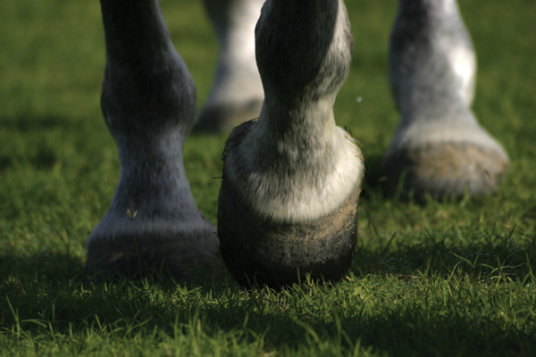
The hooves of horses are a remarkable thing. Strong and tough, yet also delicate, their health affects your entire horse’s wellbeing. That’s why it’s a good idea to keep a close eye on your horse’s feet. Craig Lesser, DVM, a podiatry veterinarian at Rood & Riddle Equine Hospital in Lexington, Ky., shares with us 5 serious hoof conditions to look out for. While some are more common than others, all require a conversation with your veterinarian for diagnosis and treatment.
Keratoma

EFFECT: This outsized tumor causes pressure necrosis, according to Lesser.
“As the keratoma grows, it puts pressure on the bone and will cause the bone to remodel,” says Lesser. “With time, it can cause lameness.”
The first sign of this hoof condition is often a chronic abscess on the affected foot or lameness.
“A lot of times, especially with a conical keratoma, you’ll see a change in the white line of the foot, which can be an indication,” says Lesser. “With a spherical one, there aren’t always changes on the outside of the sole. We usually see it as an abscess, but the actual pressure itself from bone remodeling can cause lameness.”
DIAGNOSIS: Radiographs can show the defect on the bone, and Lesser says this is the most common way veterinarians diagnose this hoof condition. They can also be diagnosed after an MRI or CT scan.
TREATMENT: Lesser says the only real option is surgical removal. Depending on the type of lesion, surgery can result in a significant layup time ranging from two or three months to up to a full year for a hoof to grow out if the lesion was removed from the coronary band.
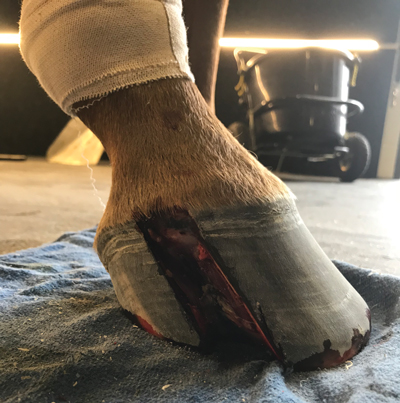
Quittor
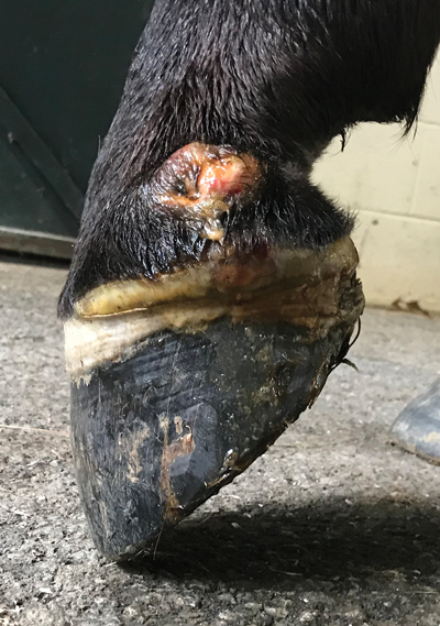
WHAT IT LOOKS LIKE: A bacterial infection that embeds behind collateral cartilage in the hoof, resulting in what looks like an abscess that has blown out of the coronary band, except it’s about a finger’s width higher than the coronary band, where the collateral cartilage ends. This cartilage is the firm-yet-elastic portion of the back half of the hoof that helps give it shape.
Lesser explains that quittor is very hard to clear up.
“It’s a tough infection to get rid of because drainage isn’t as easy as with a normal abscess,” he says.
EFFECT: The horse will usually be lame from the abscess, and once the quittor ruptures, there will be residual lameness.
DIAGNOSIS: The telltale abscess a thumbs-width above the coronary band is the first indication. X-rays with a contrast study will confirm, or an MRI will reveal the extent of the infection.
TREATMENT: A range of options are available for this hoof condition, including systemic and regional antibiotics. If the condition is bad enough, your veterinarian may put in a drain behind the collateral cartilage to allow for drainage. Lesser has also used larval therapy—incorporating fly maggots—and surgical debridement of the area as needed.
Canker
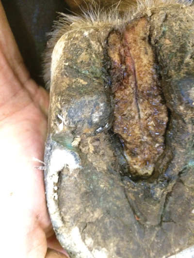
WHAT IT LOOKS LIKE: Canker looks like a really bad case of thrush at the onset.
“It forms a cauliflower-looking structure as it grows,” says Lesser. “When it gets scraped, it bleeds easily.”
It usually starts in the center of the foot and will then envelop the entire frog and sometimes the sole of the foot, in time.
“It’s a hyper-proliferation of keratin,” says Lesser.
EFFECT: Lameness is sometimes present, but if left untreated, it can cause permanent structural damage to the hoof capsule.
DIAGNOSIS: Canker is diagnosed off gross evaluation. Because the cause of the disease hasn’t been identified—it has been suggested that it comes from viruses or bacteria, and it’s also been called a cancer—it’s difficult to get a solid diagnosis.
“Canker is one of the most frustrating diseases of the foot that I deal with, because we don’t quite know what causes it,” says Lesser.
Veterinarians diagnose canker from a growth evaluation and visual examination.
TREATMENT: Lesser typically starts by blocking the hoof and doing a gross debridement to take the hoof back to healthy tissue. He will then cauterize the remaining tissue to make sure the infected tissue has been killed. He’s also used cryotherapy, laser surgical debridement, and a variety of other methods to debride the material as needed.
“The after-care is really what makes treatment successful or not,” says Lesser. “That involves months of bandaging, with or without shoes and treatment plates to make sure the disease does not come back in the future.”
Coffin Bone Infection
WHAT IT LOOKS LIKE: A coffin bone infection can affect horses of all ages. It usually starts as chronic abscesses or an abscess that has gone into the bone, causing an infection. If it worsens, it can form a sequestrum.
“A sequestrum is when a piece of the bone actually breaks off and the body starts fighting it as if it’s a foreign object,” says Lesser.
EFFECT: Severe lameness.
“It’s like an abscess that won’t go away, even if you have it open and draining,” adds Lesser.

DIAGNOSIS: Radiographs are the most common method.
TREATMENT: If the coffin bone infection creates a sequestrum, your veterinarian will surgically remove the dead piece of bone. If the coffin bone itself is affected and it’s not too bad, Lesser will treat the infection with antibiotics and larval therapy.
“If caught early and it’s fairly minor, once we get the infection under control, the horses don’t look back,” says Lesser. “But we do hit them very hard with treatment because it can be a such a bad disease.”
If a large piece of the coffin bone is removed that destabilizes the bone, it may take longer to heal. But with a smaller piece, Lesser says horses can heal nicely with no further issues.
Puncture Wound

WHAT IT LOOKS LIKE: If your horse steps on a sharp object like a nail, the nail may embed in the hoof. This is a serious injury that should be handled by your veterinarian.
EFFECT: “If the nail goes into certain structures in the hoof, it could be life-threatening for the horse,” says Lesser. “Don’t pull the nail out before the vet shows up. Make sure you call a vet right away. It’s very important.”
DIAGNOSIS: Lesser prefers to take radiographs before making a move to see what the nail is affecting inside the hoof.
TREATMENT: Lesser says the most important thing is not to pull the nail out without waiting for your veterinarian.
“If it’s sticking out of the hoof quite a bit and your horse is moving around, you can build a donut around it until your veterinarian can get out to your place,” says Lesser. You can build that donut with material at your barn—leg wraps are a great option.
If the nail isn’t touching an important structure, the veterinarian will carefully remove it, treat the horse with antibiotics and all should be well. It can be much worse—if the nail punctures the coffin bone, it can fracture the bone.

“Worst-case scenario, if it’s involving a joint or tendon sheath, those structures will need to be flushed out and [the horse given] systemic antibiotics, possibly regional limb perfusions, to treat that infection,” says Lesser.
If the puncture wound is mild, the horse could be back to work within a week. If it’s deeper or has hit a sensitive structure, it could be two to three weeks of recovery. If the nail has gotten into a joint, it could require at least a month of recovery for this hoof condition.
This article about hoof conditions appeared in the June 2020 issue of Horse Illustrated magazine. Click here to subscribe!