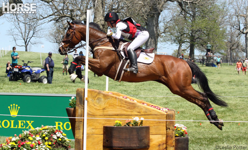
Arthritis is most common in performance horses, including eventers, jumpers, ropers, barrel racers and dressage horses.
“Healthy as a horse.” Fact or myth? For the thousands of horse owners who spend countless hours hand-walking their horse after an injury, “myth” is often the answer. But horses are essentially very healthy. It’s the work they do for us that causes most leg injuries, creating a whole world of diagnostics, treatments and prevention.
Types of Injuries
In general, exercise-related injuries can be organized into hard or soft tissue. Causes include repetitive-action injuries, which lead to the slow breakdown of tissue, and trauma/ overload injuries, which cause tearing or breaking of tissue.
By far the most common exercise-related injuries occur in soft tissues (all tissues other than bone and hoof). The most common soft-tissue injuries are in the tendons and ligaments, followed by joint capsules, joint surfaces (cartilage) and muscles. The joint capsule is the pocket around the joint that gives it structure and produces and contains the joint fluid.
 Click to view larger |
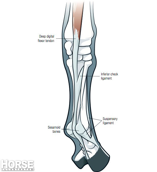
Tendons connect muscle to bone, whereas ligaments connect bone to bone. Both are made of long, thin fibers bundled together. They have incredible strength when exertional forces are applied in alignment with the fibers, but when their capacity is overloaded, or the load is not in alignment with the fibers (such as spinning and twisting on one leg), the fibers can tear. Muscles can tear or become sprained in the same fashion.
The tendons and ligaments most familiar to horse owners are the deep digital flexor and superficial flexor tendons, and the check and suspensory ligaments, all of which are located along the back of the cannon bone between the knee (or hock) and hoof. Other tendons and ligaments that might be less familiar to you include collateral ligaments (small ligaments on the inside and outside of the joint) and sesmoidian ligaments (very small ligaments criss-crossing around the sesamoid bones of the fetlock).
Understanding the Injuries
Repetitive actions or overloading a tissue may weaken it, making it susceptible to breakdown. This occurs most often on the joint surfaces and in bones. On the joint surface, the cartilage begins to break down from repetitive stress, causing the joint to become inflamed.
Once the joint is inflamed, its lubricating fluid becomes thinner, which in turn protects the joint less, allowing more cartilage to be damaged. Soon the joint capsule also becomes inflamed, developing finger-like projections (called villi) on its surface. This prohibits the joint from producing its normal thick fluid.
When combined, all of these factors produce inflammation and cartilage breakdown within the joint. When the body can no longer replace cartilage, it reacts by laying down bone on the damaged cartilage. This can be seen on radiographs, and is known as bony arthritis. This is very common in performance horses such as dressage and western pleasure mounts, jumpers, eventers, ropers, barrel racers, et cetera. It is much less common in racehorses, since their careers are much shorter.
Medical intervention can help preserve long-term joint function by reducing inflammation and stimulating physiological repair, especially when damage is caught early. Treatment options include injectables such as sodium hyaluronate (Legend), which improves the quality of synovial fluid, and polysulfated glycosaminoglycan (Adequan i.m.), which helps stimulate cartilage repair in addition to restoring synovial lubrication.
 Click to view larger |
Horses in speed disciplines incur a very different type of stress on their tissues, and therefore suffer from very different injuries. The forces created and the stresses placed on a racehorse’s legs in just one race are so tremendous that the bone density of the lower leg is reduced by about 10 percent, leaving the horse susceptible to a broken leg if he is worked too hard. Similarly, the tendons and ligaments also become weaker and can tear as a result of the forces experienced while running.
Proper Conditioning
When tissues are used regularly and their workload is gradually increased—which is the process we call conditioning—they become stronger. If the tissues are not conditioned, they are weaker and more susceptible to injury. In all disciplines, conditioning is incredibly important for prevention of exercise-induced injuries. Many injuries occur simply from the fact that the horse was too out of shape for his workload.
Do your homework on fitness and conditioning. Many factors must be considered, such as the horse’s age, breed and conformation, as well as the type of sport, et cetera. In general, a sensitive horse is very quick to give warning signs that the work is too much and a problem is brewing.
Some of the more common indicators that the tissues are weakening and injury may be imminent, if not already present, include switching leads, cross-firing, unwillingness to pick up a lead, refusing to trot or canter, bucking at the canter, heaviness on the forehand, refusing jumps, tripping, unwillingness to bend, a sore back, tossing the head, unwillingness to accept the bit, poor collection and tail swishing. These are all signs that the exercise program may be too hard for the horse and a change in training is needed.
New Technology
In the last five to 10 years, there have been many advances in the diagnosis and treatment of injuries. We can now look into the “black box” of injuries with imaging systems—such as magnetic resonance imaging (MRI), cat scans (CTs), thermography, digital radiographs (x-rays) and digital ultrasound—to actually see the tissue and the damage. Treatment methods developed in the last five years have reduced the outcome of some of these injuries from career-ending to a return to almost normal function.
The biggest breakthroughs in the treatment of these injuries have been made in the areas of regenerative medicine and energy. Regenerative medicine technology uses the body’s own tissues and chemicals to initiate healing. The two leaders in this area are stem cell therapy and chemical mediators.
• STEM CELL THERAPY: All tissue is made up of cells: bone cells, skin cells, tendon cells, et cetera. Stem cells are those that haven’t yet become any type of tissue but have the capacity to do so. This means that if stem cells are transplanted into cartilage, they become cartilage; if transplanted into tendon, they become tendon, and so on.
In relation to injuries, this means that stem cells can be placed into an injury site, where they multiply, differentiate into the tissue and regenerate the tissue, which heals with new tissue instead of scar tissue. Many tissues—such as cartilage, tendons, ligaments and nerve tissue—can’t replicate and replace themselves once damaged. They can only produce bone or scar tissue, leaving the injured site very weak with decreased capacity for work, and in many cases dysfunctional from excessive scar or bone tissue. Sometimes the damage is so severe that healing can’t occur.
Stem cells are found in bone marrow or fat. The two current treatment techniques are bone marrow transplants from the horse’s own sternum or the fat pad around the tailhead.
• CHEMICAL MEDIATORS: Chemical mediators, like stem cells, are also capable of stimulating healing. Chemical mediators are the chemical messengers within the body. They are countless in number, function and location. Our knowledge of them is limited, but what we do know has produced astonishing results.
 Click to view a chart of lower leg injuries in horses |
The leading technologies in this area are interleukin receptor antagonist protein (IRAP) and platelet-rich plasma (PRP). In both of these treatments, the horse’s blood is drawn and processed in such a way that the “good” healing chemicals are enhanced in the plasma, which is then injected into the area of injury. The chemical mediators then stimulate healing in that area. Both of these techniques have proven invaluable for injuries that occur within the joint. I have seen particular success with the use of IRAP for the treatment of collateral injuries of the coffin joint and prevention of bony arthritis in the hocks and coffin joints.
• SHOCKWAVE THERAPY: Another exciting development is in the area of energy. For years, the use of cold lasers, ultrasound, hot and cold therapies, electrostimulation and radiation have had their place in the adjunct treatment of injuries. However, none stood out as a leader for healing and regeneration until the advancement and perfection of extracorporeal shockwave units.
Extracorporeal shockwave, aka “shockwave therapy,” involves producing energy within a probe that is placed on the damaged tissue and sends energy through the injured tissue. Researchers are not yet clear on how the energy works, but they are very clear on the results: The tissues are stimulated to heal, and they heal very well. This therapy is now the forerunner in the treatment of certain soft-tissue injuries and some non-healing skin wounds and fractures.
The most common injuries treated with shockwave therapy are tendon/ligament sprains and tears, collateral ligaments, and back injuries/pain. I have seen a high percentage of return to performance in horses treated with this therapy, particularly when used on subtle sprains of the soft tissue structures around the fetlocks, hocks, stifles and back. It is considered a therapy for structures outside of the joint, and works very well in combination with regenerative therapies, such as stem cell, IRAP and PRP.
Healthy as a horse? Absolutely. Even when injuries occur, veterinarians can offer hope from the latest advancements in diagnostics and treatment. The odds are now better than ever that your horse may return to full health and his former performance level.
Liked this article? Here’s more info on soundness and lameness in horses:
New Horizons in Arthritis Treatment
The Top Five Causes of Equine Lameness
Janice Posnikoff, DVM, heads up Orange County Equine Veterinary Services in Southern California
This article originally appeared in the May 2010 issue of Horse Illustrated. Click here to subscribe.

That’s great to hear.
Wow, so many things can go wrong.