Lameness is one of the most prevalent veterinary complaints in the horse. While there are a multitude of ways horses can go lame, some conditions occur more frequently.
Lameness in the horse can range from an obvious non-weight-bearing gait to more subtle signs of discomfort that may only be displayed as poor performance. The goals of a lameness examination are to identify the affected leg and the exact part of it that is causing the lameness.

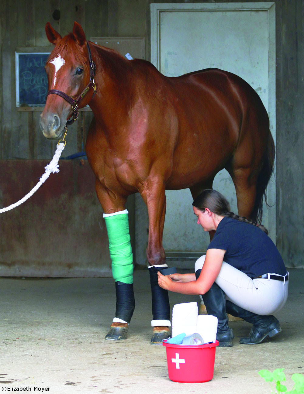
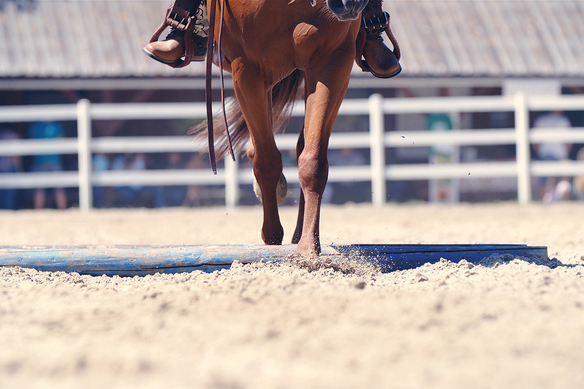
Before watching the horse move, your vet may want to know about his recent activity. The horse should be examined from a distance and up close, with a thorough palpation of the legs. As the vast majority (up to 90 percent) of all lamenesses originate in the foot, the hoof, sole and heels should be carefully assessed; applying pressure around the foot with hoof testers can help to detect any pain. Finally, the horse can be observed at the walk and trot, both in a straight line and on a circle, to look for signs of lameness.
The area of origin can be more closely isolated with flexion tests, placing pressure on a joint in the leg by flexing it; lameness often worsens after flexion.
To further pinpoint where the pain is coming from, a diagnostic nerve block can be used, where a local anesthetic is injected to numb a portion of the leg. The horse is then jogged again to see if the lameness has improved or resolved. These procedures are started at the heel/hoof and progress up the leg until the area of pain is identified.
Next, diagnostics such as radiographs (X-rays), ultrasound and even more detailed scans, including computed tomography (CT) or magnetic resonance imaging (MRI), can be employed to determine the exact cause of the lameness.
Here are the top five causes of lameness.
Foot Abscess
Subsolar abscesses, localized infections just beneath the sole of the hoof, are one of the most widespread causes of foot pain. As an abscess develops, it exerts pressure on the sensitive structures of the foot.
Continue Reading >>
Navicular Syndrome
The navicular bone is a small canoe-shaped bone that lies within the hoof behind the coffin and short pastern bones. Navicular syndrome is a term used to describe the heel pain and pathology of navicular disease.
Continue Reading >>
Heel Pain
While heel pain is a component of navicular syndrome, it is very important to point out that there are a multitude of other structures in the hoof that can cause heel pain, such as the suspensory ligament of the navicular bone, the impar ligament that connects the navicular bone to the back of the coffin bone, and other collateral ligaments.
Continue Reading >>
Degenerative Joint Disease
Degenerative joint disease (DJD) is a common occurrence in older, and sometimes not so old, equine athletes. The body is designed to maintain the cartilage in the joints, repairing damage after normal wear and tear. In athletic horses, however, excessive wear can overwhelm the repair process.
Continue Reading >>
Tendon and Ligament Injury
There are several important tendons and ligaments in the lower leg. The superficial digital flexor tendon, the deep digital flexor tendon, and the suspensory ligament are the most prominent and often prone to injury.
Continue Reading >>
While it is impossible to list all the ways why horses can go lame, it’s useful to understand some of the top causes. Knowing how these injuries and diseases present will allow you to better recognize a problem and start appropriate treatment sooner so that you can get back in the saddle.
The Lameness Scale
The American Association of Equine Practitioners has developed a grading scale (0-5) so that all horsemen and veterinarians can use the same criteria for describing a lameness.
- Grade 0: A sound horse .
- Grade 1: Lameness that is difficult to observe and is not consistently apparent.
- Grade 2: Lameness that is difficult to observe at a walk or when trotting in a straight line, but consistently apparent under certain circumstances.
- Grade 3: Lameness that is consistently observable at a trot under all circumstances.
- Grade 4: Lameness that is obvious at the walk.
- Grade 5: Lameness that produces minimal weight bearing in motion and/or at rest, or a complete inability to move.
Liked this article? Here’s more on equine lameness:
Managing Mystery Lameness
Hoof Care and Lameness Topiclist
JOAN NORTON, VMD, DACVIM, is the founder of Norton Veterinary Consulting and Education Resources, a firm dedicated to the education of horsemen and equine veterinarians through internal medicine consultations, lectures, webinars and writing. More information on her services and courses can be found at
www.nortonveterinaryconsulting.com.
This article originally appeared in the June 2014 issue of Horse Illustrated. Click here to subscribe!


The one huge consideration that was entirely missed in this otherwise detailed article is the saddle itself – which can often be the cause of misdiagnosed lameness! Check out Jochen Schleese’s book “Suffering in Silence: The Saddle Fit Link to Physical and Psychological Trauma in Horses”(available through Horsebooksetc.com and Amazon.com in e-format) for some insights into the various reasons for equine lameness. Before you go into expensive pharmaceuticals and neurological diagnoses it might be worth investing in a simple saddle fit evaluation by a qualified fitter.
Good information to know.
Slow motion video analysis is a tool that is very helpful to assess subtle lameness issues in the horse. It is an invaluable tool to a reliably assess and monitor the response to nerve and joint blocks as well as treatment and rehabilitation.