 |
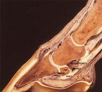
| The anatomy of the navicular bone (NB) area is quite complex with many soft tissue structures surrounding the bone including the impar and suspensory ligaments, the navicular bursa and the deep flexor tendon (DDFT). Image courtesy AAEP. |
Navicular disease was first described 250 years ago, when it was called “coffin joint lameness” because the lameness came from inside the horse’s hoof. Nearly a century later, the term “navicular joint disease” began to be used to describe the degenerative disease. Navicular affects the horse’s navicular bone, the small bone in the back of the foot, as well as associated structures such as the coffin joint, the impar ligament, suspensory ligament of the navicular bone, the navicular bursa, and the deep flexor tendon.
New technology and techniques have made it possible to visualize portions of soft tissue structures, but not until magnetic resonance imaging began to be used was the extent of these soft tissue injuries truly understood. As a result, a plethora of new information regarding injuries associated with navicular disease is now available, but the question that remains is what the new information means.
Navicular disease or navicular syndrome, which is more correctly termed palmar foot pain syndrome, accounts for one-third of all chronic forelimb lameness in horses. Why do these horses become lame? Some studies have shown that, at least for some of these animals, the interaction between the navicular bone, its bursa and deep flexor tendon creates a situation similar to osteoarthritis, except the degenerative process involves fibrocartilage on the back of the navicular bone and the adjacent flexor tendon.
Navicular disease is a degenerative disorder theoretically caused by excessive and sustained forces of compression at the distal one half of the bone. Abnormal forces from faulty conformation hypothetically cause biomechanical stresses on the navicular bone and cause remodeling changes. As long as the remodeling in response to these forces is slow, no problems occur. But when the biomechanical stresses exceed physiologic levels, degenerative disease in the fibrocartilage of the flexor surface causes changes in the underlying subchondral bone and bone marrow. Alterations in the bone lead to edema and subsequent venous hypertension and navicular bone-related pain.
Diagnosis of navicular disease is based on an overall evaluation of the hoof that includes: (1) response to diagnostic manipulative tests; (2) response of the lameness to diagnostic analgesia; (3) imaging of the foot; and (4) evaluation of hoof conformation. The diagnosis is made based on the clinical signs and may be corroborated by radiographic evaluation. However, radiographs alone do not diagnosose the disease.
The first steps developing a logical approach to the treatment of this hoof lameness are an accurate assessment of the pain and careful evaluation of hoof structure that may predispose it to the condition or cause the pain. Treatment then should be based on the type of and location of the disease. Shoeing should be the basis of all treatment, and any medicinal or surgical therapy should be as an adjunct to shoeing.
To prevent navicular disease, consistent care from a farrier is necessary to keep the hoof in good condition and to address issues of imbalance as they arise. Hoof care is also the area that needs the most research. At this point, clinical evidence of the effects of trimming and shoeing on soundness are largely empirical or the result of trial and error.
To fully understand navicular disease and injury to all the structures involved, additional research is needed to investigate the effects of trimming on the growth of hoof capsule; the effects of shoeing on the growth and physiology of the hoof capsule; and how limb and hoof conformation is related to the biomechanics of the foot and navicular bone. Studies to define how footing and type of performance affect navicular-associated structures are also needed.
By Tracy A. Turner, DVM, MS, Dipl. ACVS
Anoka Equine Veterinary Services, Elk River, MN



I hope that they can completely research this & find a completely long term forever cure. Ive known so many horses that get this & it affects thier lives forever in one way or another.
I sure hope my horse never gets this.
I hope this research gets done asap – but it also seems to me that performance horses that get pushed too far too early get this most often, and relatively early in life. Maybe that should tell us something!
My horse was recently diagnosed with Navicular Disease. Over the period of the one year that I have owned him he became increasingly irritated and fussy, had a shorter and shorter stride, could not pick up his right lead to save his life, and was reluctant to work. He did not run and play in the pasture and was just mister grumpy.
Since I did not know him in his past life as a show horse I was confused as to why his past owners kept saying he was such a hard worker and had never given them any troubles. Plus he is young and just came 7 this year.
It was not until he started limping( his right front) at the trot that I called the vet and had him checked out. Digital x-rays show degeneration along the lower edge of the Navicular Bone. I’m considering an MRI
before I do more than the corrective trimming and shoeing and the NASID’s that he is on. He seems happier and is starting to be more active in the pasture with his friends. It’s only been two weeks.
I’ve been reading online everyday about Navicular Disease trying to get a clearer picture of what is going on. Your article has been helpful in clarifying this complicated and frustrating diagnosis. From your article and the many others I have read it seems to me that the MRI along with the digital X rays are a must to really determine what part of the “Navicular” group is being affected and in the case where you know it’s the bone an MRI helps to determine if it’s just the bone or if the tendon and ligaments are involved as well.Thanks for putting articles like this out there for people like me.